Quantitative Jones matrix imaging using vectorial Fourier ptychography
Biomedical Optics Express (2022)
Xiang Dai1, Shiqi Xu1, Xi Yang1, Kevin C. Zhou1, Carolyn Glass2, Pavan Chandra Konda1, Roarke W. Horstmeyer1†
1 Department of Biomedical Engineering, Duke University, Durham, NC, USA, 27708
2 Department of Pathology, Duke University, Durham, NC, USA, 27708
Link to paper
Abstract
This paper presents a microscopic imaging technique that uses variable-angle illumination to recover the complex polarimetric properties of a specimen at high resolution and over a large field-of-view. The approach extends Fourier ptychography, which is a synthetic aperture-based imaging approach to improve resolution with phaseless measurements, to additionally account for the vectorial nature of light. After images are acquired using a standard microscope outfitted with an LED illumination array and two polarizers, our vectorial Fourier Ptychography (vFP) algorithm solves for the complex 2×2 Jones matrix of the anisotropic specimen of interest at each resolved spatial location. We introduce a new sequential Gauss-Newton-based solver that additionally jointly estimates and removes polarization-dependent imaging system aberrations. We demonstrate effective vFP performance by generating large-area (29 mm2), high-resolution (1.24 μm full-pitch) reconstructions of sample absorption, phase, orientation, diattenuation, and retardance for a variety of calibration samples and biological specimens.
Overview
In this work, we achieve complex-valued Jones matrix imaging at high resolution over a large field-of-view by using principles from Fourier ptychography (FP). Recent work has considered how FP can improve polarimetric imaging but did not demonstrate the ability to recover the specimen’s complex-valued Jones matrix, like many of the interferometric approaches above. Complex Jones matrix reconstruction offers a complete picture of the polarimetric properties of thin specimens and has been demonstrated in scanning-based ptychography, which uses a completely different measurement arrangement but is mathematically similar to FP. In this work, we build upon these prior demonstrations in ptychography to jointly reconstruct quantitative, complex-valued Jones matrix measurements at high-resolution over a large area, along with the additional benefit of computationally accounting for polarization-dependent imaging system aberrations.
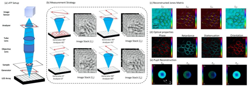
The figure above shows Overview of vectorial Fourier ptychography (vFP). (a) vFP optical setup consists of a standard polarized light microscope modified with an LED array source to sequentially illuminate specimens with light polarized by generator. Light scattered in polarization-dependent manner passes through an analyzer before being recorded by the image sensor. (b) Images are acquired for each LED under four different generator-analyzer configurations. (c) 4-channel Jones Matrix vFP reconstruction of the cross-section of a broad bean root (color shows phase). (d) Various polarization-dependent sample properties extracted from the Jones matrix using eigenvector analysis. (e) vFP simultaneously measures a polarization-dependent pupil function as it removes associated per-channel aberrations.
Results
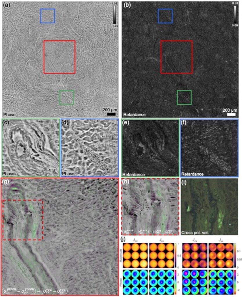
Fig. 2.Large-FOV vFP reconstruction of thin cardiac tissue section stained with Congo red dye. (a) Phase reconstruction. (b) Retardance reconstruction. (c, d) Zoom-ins of (a). (e, f) Zoom-ins of (b). (g) Difference of the difference between the real xx and yy components of the Jones matrix reconstruction between red and green channels, corresponding to the red boxes in (a) and (b). (h) Zoom-in of (g). (i) Validation via bright-field microscope imaging with crossed polarizers. (j) Spatially varying pupils, plotted as the magnitude and phase of the Jones matrix components. White dots denote the center of the pupil to improve visualization of shifts
We applied vFP to reconstruct the vectorial response of a specimen that is commonly viewed using polarization optics within the pathology lab: thin sections of fixed cardiac tissue stained with Congo red dye. Congo Red is known to stain specimen areas that contain amyloid fibrils in a manner that highlights birefringence as anomalous colors (typically, as an “apple-green” shading) when viewed between two crossed polarizers using white light illumination in a transmission geometry. Visual identification of such apple-green areas within the interstium and vessel walls of endomyocardial tissue is often an important diagnostic indicator of the presence of cardiac amyloidosis. In Fig. 2(a, b), we present a large-FOV vFP reconstruction (8.27mm2 shown) of the phase and retardance of Congo red-stained cardiac tissue, wherein the arterial wall is clearly visible. Interestingly, in Fig. 2(f), the red blood cells (small, dark circular features) located within the vessels each exhibit a higher retardance along their border, which is consistent with our observations with polystyrene beads. Examining the real component of the specimen Jones matrix entries and within this area shows minimal visual disparity un-der both green and red illumination wavelengths. However, by computing the difference ( −) for each color channel, and subtracting the red channel’s difference from the green, it is possible to produce a high-contrast map of Congo-red stained areas, as presented as a green overlay in Fig. 2(f). Comparing the resulting channel difference map to what is typically observed with polarization optics in a standard microscope (white-light illumination, NA=0.25) demonstrates a close qualitative mapping. We hypothesize that vFP can thus provide a useful tool for examining the polarimetric response of such Congo-red-stained cardiac tissue over large areas at high resolution. Reconstructing a large FOV with vFP additionally allows us to examine how the recovered Jones matrix of the imaging system pupil function differs between on-axis and off-axis sample positions (Fig. 2(j)). Here, we divided the reconstruction FOV into 3×3 regions and averaged all pupils within each region. The Jones matrix pupil phase patterns experience a shift from the center for off-axis locations. Interestingly, the and pupil amplitudes are spatially varying, unlike and . Higher-NA objectives or cheaper plastic lenses may exhibit stronger polarization effects, which will be the subject of future investigation