Uncategorized

scatterBrains: an open database of human head models and companion optode locations for realistic Monte Carlo photon simulations

scatterBrains: an open database of human head models and companion optode locations for realistic Monte Carlo photon simulations Melissa M. Wu1,2, Roarke Horstmeyer1, Stefan A. Carp2 1Department of Biomedical Engineering, Duke University, Durham NC, USA. 2 Athinoula A. Martinos Center for Biomedical Imaging, Charlestown, MA, USA. Link to paper and code Abstract Monte Carlo (MC) …
scatterBrains: an open database of human head models and companion optode locations for realistic Monte Carlo photon simulations Read More »

Imaging Dynamics Beneath Turbid Media via Parallelized Single-Photon Detection

Link to paper Summary ________________________________________ Noninvasive optical imaging through dynamic scattering media has numerous important biomedical applications but still remains a challenging task. While standard diffuse imaging methods measure optical absorption or fluorescent emission, it is also well-established that the temporal correlation of scattered coherent light diffuses through tissue much like optical intensity. Few works …

Imaging Dynamics Beneath Turbid Media via Parallelized Single-Photon Detection Read More »

Tensorial tomographic Fourier ptychography with applications to muscle tissue imaging

Link to paper Summary ________________________________________ We report tensorial tomographic Fourier ptychography (T2oFu), a nonscanning label-free tomographic microscopy method for simultaneous imaging of quantitative phase and anisotropic specimen information in 3D. Built upon Fourier ptychography, a quantitative phase imaging technique, T2oFu additionally highlights the vectorial nature of light. The imaging setup consists of a standard microscope …

Tensorial tomographic Fourier ptychography with applications to muscle tissue imaging Read More »

Review on Digital staining in Optical Microscopy using Deep Learning

Our review on Digital staining in optical microscopy using deep learning ________________________________________ Lucas Kreiss1,2, Shaowei Jiang3 , Xiang Li4, Shiqi Xu1, Kevin C. Zhou1,5, Kyung Chul Lee1,6, Alexander Mühlberg2, Kanghyun Kim1, Amey Chaware1, Michael Ando7, Laura Barisoni8, Seung Ah Lee6, Guoan Zheng3, Kyle J. Lafata4, Oliver Friedrich 2 and Roarke Horstmeyer1 1 Department of Biomedical …

Review on Digital staining in Optical Microscopy using Deep Learning Read More »

Quantitative Jones matrix imaging using vectorial Fourier ptychography

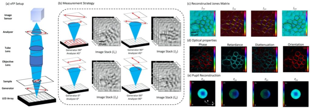
Quantitative Jones matrix imaging using vectorial Fourier ptychography Biomedical Optics Express(2022) Xiang Dai1, Shiqi Xu1, Xi Yang1, Kevin C. Zhou1, Carolyn Glass2, Pavan Chandra Konda1, Roarke W. Horstmeyer1† 1 Department of Biomedical Engineering, Duke University, Durham, NC, USA, 27708 2 Department of Pathology, Duke University, Durham, NC, USA, 27708 Link to paper Abstract This paper …

Quantitative Jones matrix imaging using vectorial Fourier ptychography Read More »

Vectorial Fourier ptychography

Quantitative Jones matrix imaging using vectorial Fourier ptychography Biomedical Optics Express (2022) Xiang Dai1, Shiqi Xu1, Xi Yang1, Kevin C. Zhou1, Carolyn Glass2, Pavan Chandra Konda1, Roarke W. Horstmeyer1† 1 Department of Biomedical Engineering, Duke University, Durham, NC, USA, 27708 2 Department of Pathology, Duke University, Durham, NC, USA, 27708 Link to paper Abstract This …

Vectorial Fourier ptychography Read More »

Quantized FP with binary images from SPAD Cameras

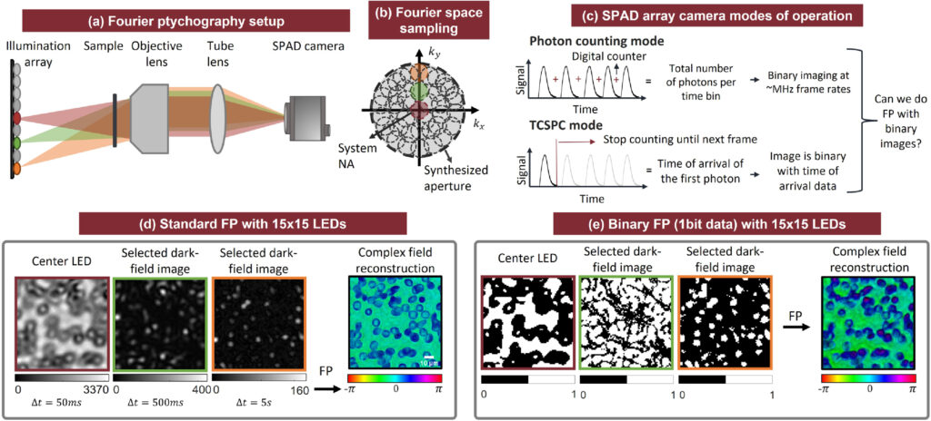
Quantized Fourier ptychography with binary images from SPAD cameras Paper Published in Photonics Research(2021) Xi Yang,1,* Pavan Chandra Konda,1 Shiqi Xu,1 Liheng Bian,2 and Roarke Horstmeyer1,3,4 1Department of Biomedical Engineering, Duke University, Durham, North Carolina 27708, USA 2School of Information and Electronics, Beijing Institute of Technology, Beijing 100081, China 3Department of Physics, Duke University, Durham, North …

Quantized FP with binary images from SPAD Cameras Read More »

Deep Optical Blood Analysis

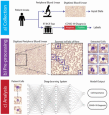
Deep Optical Blood Analysis: COVID-19 Detection as a Case Study in Next Generation Blood Screening medRxiv(2021) Authors: Colin L. Cooke, Kanghyun Kim, Shiqi Xu, Amey Chaware, Xing Yao, Xi Yang, Jadee Neff, Patricia Pittman, Chad McCall, Carolyn Glass, Xiaoyin Sara Jiang, Roarke Horstmeyer Code Paper Abstract Visual examination of cell morphology within a peripheral blood …

Deep Optical Blood Analysis Read More »

Learned Sensing

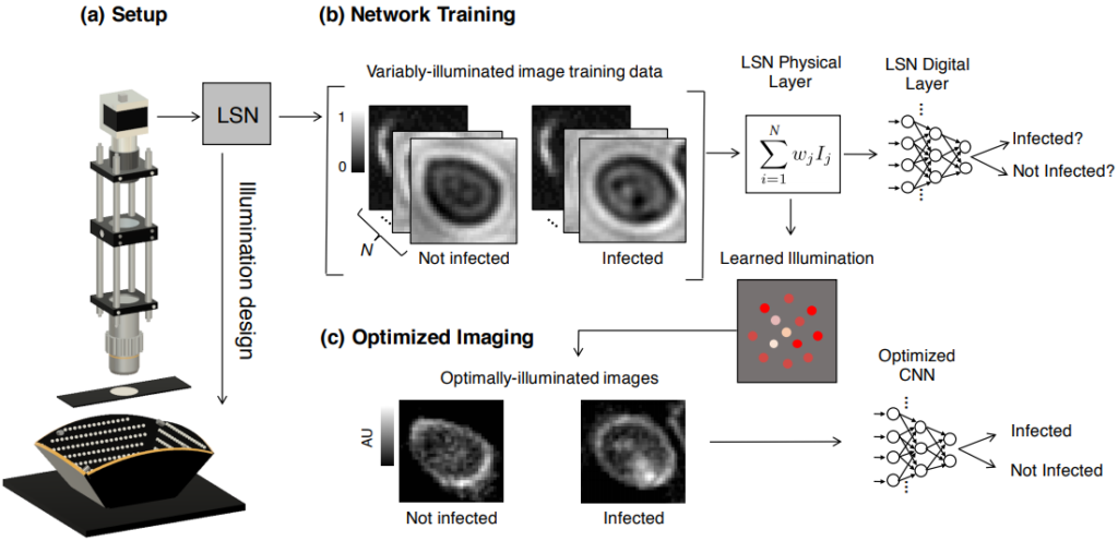
Learned Sensing: Jointly optimized microscope hardware for accurate image classification Biomedical Optics Express (2019) Alex Muthumbi1,+,Amey Chaware2,+, Kanghyun Kim2, Kevin Zhou3, Pavan Chandra Konda3, Richard Chen4, Benjamin Judkewitz5, Andreas Erdmann6,1, Barbara Kappes7, Roarke Horstmeyer2,3 1: School of Advanced Optical Technologies, Friedrich-Alexander University, Erlangen, Germany. 2: Department of Electrical and Computer Engineering, Duke University, Durham NC, …

Learned Sensing Read More »

// First try loading jQuery from Google's CDN